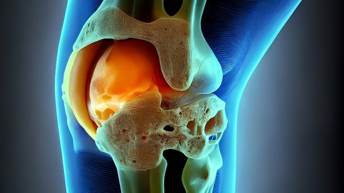
(HealthDay News) — (Tasrir) — Michelle Villagran, from Wellesley College in Massachusetts, and colleagues determined the optimal meniscal radiomic features for classifying people who will develop an incident destabilizing medial meniscal tear. Magnetic resonance images from an existing case-control study that included images from the first four years of the Osteoarthritis Initiative (OAI) were analyzed; the sample was limited to 215 patients. At the OAI baseline visit, one reader manually segmented each participant’s anterior and posterior horn of the medial menisci. From each medial meniscus horn, 61 different radiomic features were extracted. To determine the classification rules and important variables that predict an incident destabilizing meniscal tear, a classification and regression tree (CART) analysis was performed.
The researchers found that the CART correctly classified 24 of 34 cases and 172 of 181 controls, with sensitivity and specificity of 70.6 and 95.0 percent, respectively. Large zone high gray level emphasis from the posterior horn was identified by CART as the most important variable to classify who would develop an incident destabilizing medial meniscal tear.
“The use of radiomic features provides more sensitive and quantitative measures of meniscal alterations, allowing us to intervene and prevent devastating meniscal tears,” the authors write.
The study was partially funded by Merck, Novartis, GlaxoSmithKline, and Pfizer.


